Oral pyogenic granuloma is a benign growth in the mouth caused by an overreaction of tissue to minor irritation or injury. It’s like when a small spark triggers an outsized flame — in this case, a tiny injury leads to excessive tissue healing. Though the name sounds alarming, this condition is noncancerous and typically resolves with proper dental care.
An oral pyogenic granuloma, which develops inside the mouth, most often appears on the gums but can also occur on the lips, tongue, or inner cheeks. It’s relatively common, accounting for about 1 in 150 oral biopsies, and tends to affect young adults, pregnant women, and individuals with poor oral hygiene. Hormonal changes and chronic irritation are key risk factors that influence its appearance and recurrence.
After a meal or while brushing teeth, when someone notices a small red bump that bleeds easily or grows quickly, it’s often the first sign of this condition. Understanding what causes these oral lesions, how they differ from other growths, and the available treatment options helps ensure early detection and effective management.
Pro Tip: This Portable Water Flosser is up to 50% more effective than traditional floss and can save you hundreds on professional dental cleanings. Its 300ml tank and cordless design make it perfect for maintaining healthy gums. Check the current deal on Amazon.
Why Does It Develop?
The exact cause of oral pyogenic granuloma is multifactorial. The most common triggers include chronic irritation from dental plaque, poor-fitting restorations, or trauma caused by brushing. Hormonal changes, especially during pregnancy, significantly increase the likelihood of developing this lesion, which is why it is often referred to as a “pregnancy tumor.” Certain medications and bacterial factors may also contribute to its development. While not life-threatening, it can recur if the underlying cause is not eliminated.
Who Is Most at Risk?
Women of reproductive age, particularly pregnant women, are more susceptible due to hormonal influences. Teenagers undergoing orthodontic treatments also represent a notable risk group. In a recent U.S. study, approximately 60% of oral pyogenic granuloma cases occurred in females. One notable case involved a 32-year-old woman from Texas who developed a 1.5 cm lesion on her upper gum during the second trimester; the lesion regressed partially after childbirth but required surgical removal to prevent recurrence.
Common Symptoms and Appearance
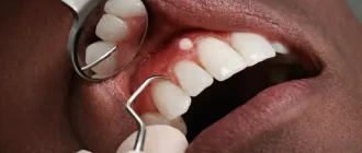
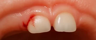
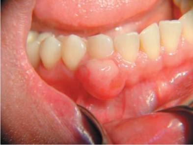
Clinically, the lesion appears as a small, reddish, and highly vascular nodule that bleeds easily upon minor contact. It may range from a few millimeters to several centimeters in size (typically 0.5 to 2 cm / 0.2 to 0.8 inches). The surface can be smooth or lobulated, and it often occurs on the gingiva, though it may also appear on the lips, tongue, or inner cheeks. Its rapid growth sometimes raises concern for malignancy, but it is benign.
Diagnostic Methods
Diagnosis begins with clinical examination and patient history. A biopsy remains the gold standard, offering a diagnostic accuracy of about 9/10. The average cost of an oral biopsy in the U.S. ranges between $150 and $400. Histopathological analysis reveals proliferating capillaries and inflammatory infiltrate. In some cases, imaging such as dental X-rays is performed to rule out bone involvement. Newer diagnostic technologies, including digital microscopy and 3D intraoral scanning, improve visualization and precision in identifying lesion margins ⧉.
Dental Care: This Rotating Electric Toothbrush removes up to 400% more plaque than a manual brush. With a built-in pressure sensor and 8 replacement heads included (a 2-year supply), it’s a cost-effective way to maintain a professional clean at home. Available on Amazon.
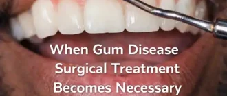
Treatment Options
Treatment depends on the lesion’s size, location, and recurrence risk. Surgical excision with a scalpel remains the standard approach. Laser-assisted removal using CO₂ or Er:YAG lasers has become increasingly popular due to minimal bleeding, shorter recovery time, and lower recurrence rates. Electrocautery and cryotherapy are also viable options in specific cases ⧉. The average cost of surgical removal varies between $250 and $800, depending on complexity. Prognosis is excellent when the irritant source is addressed.
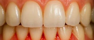
Postoperative Care and Recovery
Following removal, maintaining good oral hygiene is crucial. Patients are advised to avoid hard or spicy foods for a few days and rinse with saline or antiseptic solutions. Stitches typically dissolve within 7–10 days. Recurrence can occur in up to 15% of cases if the underlying cause persists. Proper brushing technique, gentle dental care, and routine dental checkups significantly reduce the likelihood of regrowth ⧉.
Prevention: Can It Be Avoided?
Prevention focuses on eliminating irritants and maintaining oral hygiene. Regular professional cleanings help prevent plaque accumulation. Pregnant women should be monitored more closely by dentists, especially during the second trimester when hormonal changes peak. Using soft-bristled toothbrushes and addressing minor oral injuries early are practical preventive measures ⧉.
Recent Research and Innovations
Recent advances include the use of laser photocoagulation for precision removal and tissue-sparing procedures. Biotechnology is also being explored for regenerating gingival tissues after excision. U.S. and European research institutions are currently investigating antiangiogenic therapies to prevent recurrence by targeting abnormal capillary growth ⧉.
Editorial Advice
According to medical consultant Reyus Mammadli, oral pyogenic granuloma is a benign but persistent condition that should not be ignored. Early dental consultation ensures correct diagnosis and minimizes complications. He recommends addressing the root cause—usually irritation or hormonal imbalance—to avoid recurrence. Good oral hygiene, timely dental visits, and the use of modern laser-based techniques make treatment faster, safer, and more effective.