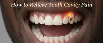
Have you ever wondered why your dentist insists on taking X-rays of your teeth, even when everything seems fine? Dental X-rays are more than just routine checks; they’re essential tools in modern dentistry.
Dental Issues Detected by X-Rays
| Issue | Percentage of Detection |
|---|---|
| Cavities | 40% |
| Bone Loss | 25% |
| Impacted Teeth | 20% |
| Infections | 15% |
What Are Dental X-Rays, and Why Are They Used?
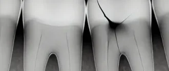
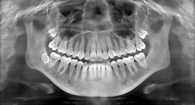
Dental X-rays, also known as radiographs, are specialized images of your teeth, gums, and jawbone. They help dentists detect issues that are not visible during a regular dental exam. These include:
- Cavities: Tiny holes or decay between teeth that are invisible to the naked eye.
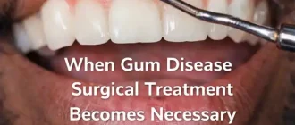
- Bone Loss: Indicators of gum disease or other jawbone conditions.
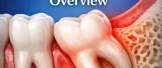
- Impacted Teeth: Teeth that have not erupted properly, often wisdom teeth.
- Infections or Abscesses: Hidden pockets of infection at the root of a tooth.
- Oral Tumors or Cysts: Rare but serious conditions that may be caught early.
Using these images, dentists can develop a personalized treatment plan to address both current and potential issues.
Pro Tip: This Portable Water Flosser is up to 50% more effective than traditional floss and can save you hundreds on professional dental cleanings. Its 300ml tank and cordless design make it perfect for maintaining healthy gums. Check the current deal on Amazon.
How Do Dental X-Rays Work?
X-rays use a small dose of radiation to create images of the internal structures of the teeth and jaw. Modern X-ray technology, including digital radiography, significantly reduces exposure compared to traditional film X-rays.
Digital X-rays work by capturing the image electronically and displaying it instantly on a monitor. This advancement not only improves accuracy but also allows dentists to enhance and zoom in on specific areas for a detailed analysis.
Are Dental X-Rays Safe?
Yes, dental X-rays are extremely safe. Modern advancements have minimized radiation exposure, making it comparable to the radiation you’d experience during a short airplane flight. Dentists also use lead aprons and thyroid collars to further protect sensitive areas.
Dental Care: This Rotating Electric Toothbrush removes up to 400% more plaque than a manual brush. With a built-in pressure sensor and 8 replacement heads included (a 2-year supply), it’s a cost-effective way to maintain a professional clean at home. Available on Amazon.
To put it into perspective:
- Bitewing X-rays: Approx. 0.005 millisieverts (mSv)
- Panoramic X-rays: Approx. 0.01 mSv
For comparison, a chest X-ray exposes you to 0.1 mSv. It’s a fraction of the exposure you’d naturally receive from the environment annually (~3 mSv).
How Often Do You Need Dental X-Rays?
The frequency depends on your age, oral health, and risk factors. Here’s a general guide:
- Children: Every 6-12 months, as their teeth and jaws are still developing.
- Adults with Good Oral Health: Every 2-3 years.
- High-Risk Patients: More frequently, especially if you have a history of cavities, gum disease, or other dental issues.
Did You Know?
- X-rays were discovered by accident: Wilhelm Röntgen discovered X-rays in 1895 while experimenting with cathode rays. (Source: National Institute of Health)
- Dental X-rays have evolved: The first dental X-ray was taken in 1896, exposing patients to dangerous levels of radiation compared to today’s minimal exposure.
Modern Trends in Dental Imaging
The field of dental imaging is rapidly advancing. Here are some trends:
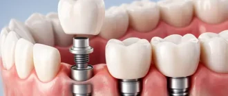
- 3D Imaging: Cone Beam Computed Tomography (CBCT) provides a 3D view of teeth, nerves, and jawbones, useful for complex cases like dental implants.
- AI Integration: Artificial intelligence is being used to analyze X-ray images, identifying potential issues with greater accuracy.
- Eco-Friendly Practices: Digital X-rays reduce chemical waste from traditional film processing.
Evolution of X-Ray Technology
| Year | Milestone |
|---|---|
| 1895 | Discovery of X-rays by Wilhelm Röntgen |
| 1896 | First dental X-ray performed |
| 1955 | Introduction of panoramic X-rays |
| 1980s | Digital X-ray technology developed |
| 2000s | 3D Cone Beam Computed Tomography (CBCT) introduced |
| 2020s | AI integration into dental X-ray diagnostics |
Are There Alternatives to X-Rays?
While there are no direct replacements, emerging technologies aim to complement or reduce reliance on X-rays. For instance:
- Near-Infrared Imaging: A non-radiative technology that shows cavities in enamel.
- Ultrasound Scans: Used experimentally for imaging soft tissues of the mouth.
Common Concerns About X-Rays
“Can X-rays harm my unborn baby?”
Dental X-rays are generally safe during pregnancy when necessary. Dentists take extra precautions, such as using a lead apron, to minimize any risk.
“Can I refuse X-rays?”
Yes, but it’s not recommended. Skipping X-rays can result in undiagnosed problems that may worsen over time, leading to more invasive and costly treatments.
Editorial Advice
Regular dental X-rays are not just about diagnosing problems; they’re about prevention and precision. By identifying issues early, you can avoid more extensive treatments and ensure better oral health. Always discuss with your dentist if you have concerns about radiation exposure or the necessity of X-rays for your specific situation. Remember, when it comes to your teeth, an ounce of prevention is worth a pound of cure.