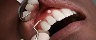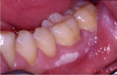
Leukoplakia, also known as oral leukoplakia, is a condition where thick, white patches develop on the tongue, gums, and inner cheeks. These patches cannot be easily removed by scraping. The sores may vary in appearance, but they are typically white or gray, thick, and slightly raised with a firm texture.
Leukoplakia typically does not cause pain, although some people may experience sensitivity in the affected area. The patches often develop slowly over a period of weeks or months and can have a smooth or slightly rough texture, as well as being either flat or raised.
There is a belief that the disease may originate from continuous irritation, although this is not always true. While eliminating the cause of irritation can help relieve the problem, in some situations, surgical removal of the sores may be necessary.
Pro Tip: This Portable Water Flosser is up to 50% more effective than traditional floss and can save you hundreds on professional dental cleanings. Its 300ml tank and cordless design make it perfect for maintaining healthy gums. Check the current deal on Amazon.
The main reason for the increase of tobacco growth in the mouth is believed to be its usage. Smokers are six times more likely to experience this compared to non-smokers. Additionally, it could be linked to the consumption of alcohol and betel quid.
In most cases, leukoplakia is not harmful and only affects the appearance. However, there are instances when leukoplakia may indicate the development of oral cancer or malignancy. It is important to handle any oral lesions with caution until they are confirmed to be non-cancerous.
What is the main cause of leukoplakia?
Here are a few of the most prevalent reasons for the occurrence of leukoplakia:
- Discomfort caused by rough teeth or dental fillings.
- Habitual cheek or lip biting
- Ill-fitting dentures
- the use of tobacco, whether through smoking or chewing
- Sunlight exposure
- AIDS or HIV
- Candidiasis
- Lupus erythematosus
What are the types of leukoplakia?
There are several classifications of leukoplakia, such as:
Dental Care: This Rotating Electric Toothbrush removes up to 400% more plaque than a manual brush. With a built-in pressure sensor and 8 replacement heads included (a 2-year supply), it’s a cost-effective way to maintain a professional clean at home. Available on Amazon.
Homogenous leukoplakia
- All white abnormalities without any indication of red.
- Oral or mouth cancer poses a very minimal risk, and it is uncommon for the cells in a uniform lesion to be precancerous.
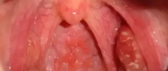
Nonhomogenous leukoplakia
- A combination of white and red spots that is not evenly distributed.
- If the red regions are not addressed, there is a higher possibility for them to develop into precancerous or cancerous conditions.
Proliferative verrucous leukoplakia
- A rare form of white patches, primarily found in older women.
- Lesions can be found throughout the entirety of the mouth rather than being confined to specific areas like the cheek and tongue.
- Tongue cancer, as well as oral cancer, can be caused by this.
Hairy leukoplakia
- Hairy leukoplakia is characterized by lesions that have folds or ridges, and these lesions also have thin hair-like filaments growing from them.
- The outcomes that occur when the immune system is weakened after being targeted by the Epstein-Barr virus and HIV.
What are the indications and manifestations of leukoplakia?
The signs of leukoplakia can resemble symptoms of other diseases, such as the following:
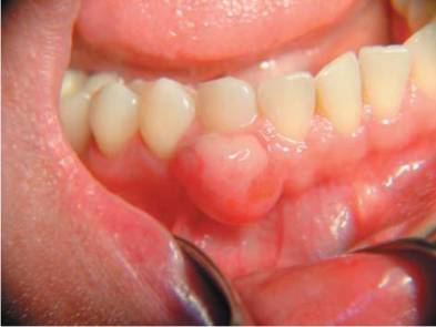
- Leukoplakia is primarily characterized by raised or toughened white patches in the mouth, specifically on the inner cheeks or tongue.
- Typically, these patches or sores are not harmful and individuals usually do not notice them until they become noticeable.
- In the mouth, one might observe red scaly patches or a mixture of raised pimples that are both red and white in color.
- These rough-looking white or red and white patches are easily identified due to their distinct characteristic of not dissolving when the mouth is cleaned or scraped using the appropriate dental tool.
How is leukoplakia diagnosed?
Your healthcare provider will examine your mouth and try to remove the white areas. If the white patches can be easily wiped off, it is likely not leukoplakia. They will also ask about your medical history, including any habits like smoking or chewing tobacco, to identify potential risk factors.
Determining whether the spots are leukoplakia or oral thrush can sometimes be difficult simply by observing them. In order to identify the cause of the patches, a doctor will have to perform certain tests.
It is possible that you will undergo cancer screening, during which the doctor will examine for any potential indicators and recommend a biopsy.
A biopsy is a procedure where a small piece of tissue is taken from the body to examine it for diseases such as cancer.
Tests for cancer screening in individuals with leukoplakia may include the following:
Oral brush biopsy
- A tiny rotating brush is utilized to eliminate certain cells from the exterior area of the abnormality.
- The cells sample that has been gathered is then examined to determine if it is cancerous.
Excisional biopsy
- Doctors surgically remove a portion of tissue from the leukoplakia lesion and evaluate it for signs of cancer.
- The use of excisional biopsy is a more accurate method for identifying cancer compared to an oral brush biopsy.
If the results of the excisional biopsy show cancer, your doctor may recommend you to a specialist for additional treatment. Sometimes, the leukoplakia patch may be very small and can be completely removed with the excisional biopsy. This means that no further treatment may be needed, and you will only need to be monitored regularly.
How do you get rid of leukoplakia?
Early diagnosis and treatment is crucial for effectively managing leukoplakia. Mild cases do not need proactive treatment and typically resolve on their own. However, if treatment is required, the first step should be eliminating the underlying cause or source of irritation.
If leukoplakia develops due to a tooth being rough or an uneven surface on a denture or filling, the dentist will treat it by smoothing the tooth and adjusting the dental equipment.
If it is determined that smoking is the reason behind your leukoplakia, you will be advised to decrease or quit smoking altogether, or consider using alternative tobacco products.
Leukoplakia is typically not harmful, and the lesions usually go away within a few weeks or months after the source of irritation has been eliminated. However, if the irritation persists and the leukoplakia does not improve, it may be necessary to surgically remove the lesion. This can be done by either a regular dentist or an oral surgeon.
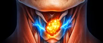
Is leukoplakia cancerous?
While not all instances of leukoplakia result in cancer, oral leukoplakia has the potential to become cancerous and is commonly observed in individuals who smoke.
Individuals who have oral leukoplakia are at a higher risk of developing oral cancer compared to those with healthy oral mucosa. Leukoplakia is a commonly observed potentially harmful oral condition, affecting 1.5 to 2.6 percent of the global population.
Early identification, management, and observation are essential due to the variable progression rates, between 0.1 to 17.5 percent, of this precancerous state to squamous cell carcinoma, a type of oral cancer.
The main goal when dealing with oral leukoplakia is to stop it from developing into invasive cancer. This can be achieved through different treatment methods like traditional knife removal, cryotherapy, and carbon dioxide laser therapy.
How can you prevent leukoplakia?
Preventing leukoplakia is the most effective way to treat it. The following recommendations can help you avoid developing these abnormal patches:
- Stop smoking and using tobacco products.
- Quitting alcohol consumption.
- It is recommended to visit the dentist twice annually for regular check-ups.