X-ray diagnosis is the easiest and most inexpensive method of examination in dentistry. It is indispensable for detecting hidden pathologies that are localized under the gums, in the jawbone or dental roots. X-rays are also used to monitor the results of therapy at every stage.
X-rays are universally used in dentistry – they are one of the indispensable prerequisites for the diagnostic examination in most clinical cases. Visual examination cannot provide detailed information about the current situation, identify hidden pathologies and give the most accurate assessment of the extent of the existing problem – in these cases, X-rays come to the rescue. This diagnostic method is also necessarily used before orthodontic treatment, prosthetics, implantation and surgery – it helps to examine the structure of the dentoalveolar apparatus in detail, assess the condition of the bone tissue and mucosa, and discover any hidden contraindications.
When X-rays are appointed
Many dental problems cannot be detected by visual examination alone. It is also necessary to consider that some pathologies are latent and do not make themselves known for a long time. This is the answer to the question of why do x-ray in dentistry, and what does the picture show – the image provides more detailed information on the current state of tissues of the mouth and teeth, can identify hidden pathological processes, to detect possible contraindications to the upcoming treatment.
Pro Tip: This Portable Water Flosser is up to 50% more effective than traditional floss and can save you hundreds on professional dental cleanings. Its 300ml tank and cordless design make it perfect for maintaining healthy gums. Check the current deal on Amazon.
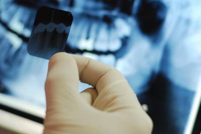
A picture is taken with a special radiographic unit, which allows the specialist to examine the clinical picture in more detail, make a correct diagnosis and prescribe an appropriate treatment. The results of such an examination provide the following possibilities:
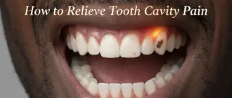
- Detect hidden injuries to the tooth, such as a crack or root fracture,
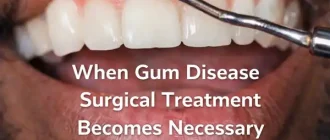
- Determine the degree of involvement of soft tissues by periodontitis and periodontal disease,
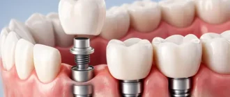
- assess the volume, height and condition of the bone prior to implantation,
- identify hidden foci of inflammation, neoplasms in bone and soft tissue,
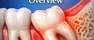
- assess the correctness of occlusion, diagnose anomalies of the dentoalveolar apparatus, detect retained elements, including the position of wisdom teeth – with all these nuances to prescribe the necessary orthodontic treatment.
Therefore, X-rays are a must before placing braces or any other corrective system, before treating dental canals, prosthetics, implants, and bone grafting. In some cases, a repeat procedure is prescribed after treatment to assess the results achieved.
Types of X-rays used in dentistry
Today, various types of radiographs are used in dental practice. Let’s look at the possible options in a little more detail:
- Targeted image: an image of 1-2 teeth, which is used to study the dentin tissues, dental root and canals, the condition of the bone tissue and the adjacent gum. This type of X-ray also allows detecting early caries, the first signs of periodontal disease, which is often asymptomatic for the patient,
- Interproximal X-ray: used primarily for examination of the crown portion of the tooth, allows the detection of hidden carious cavities, including those on the neighboring teeth of the defect and in the cervical area, makes it possible to assess the extent of the destruction of hard tissue, to detect pathological processes under crowns and fillings. It may take 3-4 pictures to get the required result,
- occlusal: the technique enables to assess the condition of the hard palate, detect neoplasms, examine the condition of submandibular and submandibular glands. The image also provides detailed information on the retained and distorted teeth, and this method is used when the aim X-ray is inconclusive for some reason,
- Long-focus X-ray: allows to closely study the periodontal tissues, that is why it is widely used in periodontology. A more detailed picture can be obtained by using a diagnostic unit with a more powerful X-ray tube,
- Panoramic picture (orthopantomogram): images of both jaws, with the possibility to examine thoroughly the crown and root of each tooth, and the periodontal tissues. This examination takes place before orthodontic treatment, prosthetics, as a part of implantation planning. The imaging also provides an opportunity to recognize buried neoplasms, trauma and other deformities of the jaw apparatus.
Separately, we can highlight computed tomography, a technique that allows you to get the most informative 3D-image with the ability to study the condition of soft and hard tissues in layers of the oral cavity. It is a more advanced technology that is necessarily used today before complicated treatment, surgery and dental implantation. It allows us to study the clinical picture in detail, identify hidden pathologies and rule out possible contraindications to the upcoming treatment.
Dental Care: This Rotating Electric Toothbrush removes up to 400% more plaque than a manual brush. With a built-in pressure sensor and 8 replacement heads included (a 2-year supply), it’s a cost-effective way to maintain a professional clean at home. Available on Amazon.
Examination methods and permissible radiation doses
Several basic methods of radiographic examination are used in dentistry. The choice of a particular diagnostic option is made by the attending physician, based on the characteristics of the clinical picture and the goals set. The radiation dose varies in each case, but is always minimal and completely safe for health:
- a targeted image – 5 μSv,
- Orthopantomogram (panoramic image) – 35 μSv,
- CT scan of both jaws (3D image) – 60 µSv1.
In order to understand whether such examinations are harmful, pay attention to the following figures: the maximum permitted annual radiation exposure for adults is 1000 µSv, and 300-400 µSv for children up to 15 years of age.
It should also be noted that before dental implantation, especially before applying one-step treatment protocols, it is necessary to get as detailed and informative scans as possible. It is important to give an accurate assessment of the condition of the bone tissue and mucosa, to study the peculiarities of the structure of the jaw apparatus, to identify possible contraindications. For this, patients are usually referred to specialized diagnostic centers, which have higher quality and more accurate equipment than private dental clinics.
How often the procedure can be repeated
Many patients are concerned about whether X-rays are dangerous and how many times they can be taken. The permissible frequency of the procedure will depend on the type of machine used. As mentioned above, the maximum radiation exposure for an adult corresponds to 1,000 μSv per year, and for children under 15 years of age – 300-400 μSv. Let’s see below what number of x-rays is acceptable without danger to health.
For adults
The maximum allowable number of scans on a radiovisiograph is 500, and on an x-ray machine is 100. Obviously, it makes no sense to undergo X-ray examination so often, no matter how complicated and time-consuming the treatment. So there is no need to worry about harm from such diagnostics.
For the child
Many parents worry about whether or not it is harmful to undergo X-rays for children. It should be said right away that there is no serious danger from such procedures. But the method allows you to identify hidden cavities, to study the process of formation of the dentoalveolar system, and if necessary, prescribe orthodontic treatment.
Let us remind that the annual allowable dose for a child is 300-400 mcSv, while modern diagnostic equipment gives a negligible dose of radiation, absolutely safe for the health of the child’s body. A special apron will be put on the child before the procedure to further reduce the degree of negative exposure.
X-ray diagnosis during pregnancy and lactation
Now let’s move on to the question of whether it is possible to take an X-ray during pregnancy and breastfeeding. Here it is important to consider the exact term, since this condition is a relative contraindication. So, in the first trimester you can not undergo X-ray diagnosis – this is a critical period, during which all the vital organs of the child are formed. This procedure is also not recommended in the last trimester, and the optimal time is the second trimester.
During lactation you can undergo X-rays, and the main thing here – to protect the chest with a special lead apron, which will give you a specialist. Answering the question about whether you can feed after the procedure, the experts disagree. To avoid any risks, it is better to express milk and for the next 48 hours to transfer the child to alternative feeding.
Dental X-rays during pregnancy: important nuances
X-rays are used with limitations in pregnancy. Pregnancy is a relative contraindication to X-rays. Therefore, the patient should warn the doctor about her situation and name the exact date of pregnancy.
Important nuances for expectant mothers:
- It is strictly forbidden to take X-rays in the first trimester, because during this period all the vital organs of the child are formed;
- The safest type of X-ray diagnosis is an orthopantomogram (panoramic image), which has a lower radiation exposure;
- If possible, it is better to use alternative methods, such as computerized visiography.
X-rays are allowed for breastfeeding, but the main thing is to cover the chest with a protective lead apron, which the doctor will give you. Pumping milk after the procedure is not necessary.
Can X-rays be taken of children’s teeth
X-rays are also widely used in pediatric dentistry. Such diagnosis is indicated for both permanent and deciduous teeth.
- With the help of X-rays, it is possible to:
- Detect various abnormalities in the eruption of crowns;
- Plan orthodontic treatment;
- determine the method of treatment for caries or pulpitis of temporary teeth, because their roots are prone to resorption (resorption).
The dangers of undergoing X-rays in dentistry
We can talk about a threat to the health of the patient only if there are violations of the technology of the procedure or exceeding the permissible standards of radiation. Among the possible negative reactions in such situations, experts highlight the following phenomena:
- changes in the composition of protein and blood,
- ionization of molecules,
- disruptions in the production of new cells,
- acceleration of aging processes.
It should be noted that the risk of encountering such troubles is extremely small. In any case, to minimize the negative impact on the body, you should wear a special lead apron and not expose yourself to X-rays too often.
Contraindications to the procedure
Sometimes due to the loss of contrast the results of X-ray diagnosis may be distorted or not informative at all. This may be due to the following reasons:
- The presence of cysts in the examined area,
- incorrectly performed fillings,
- presence of materials that shine on the X-ray image,
- Cementoma development – a tumor that occurs in the area of dental roots and resembles the cementum of a tooth.
Also, purulent inflammation can affect the quality of the final images, and some difficulties are noted in the study of wisdom teeth. The procedure is not recommended for women in the first and last trimester of pregnancy. But in general, it is the most informative and safe option for diagnostic examination.
Are dental X-rays harmful for teeth?
The maximum allowable dose of X-rays is 1000 μSv per year, which is about 100 shots.
Therefore, you should not worry if you were prescribed only 2 procedures (before and after treatment), it will not cause significant harm to the body. The radiation dose will be as negligible as in daily contact with household appliances or during an air flight to a neighboring country.
The degree of radiation exposure also depends on the novelty of the X-ray machine, so it is better to go to clinics equipped with the latest technology.
How to prepare for the examination
No special preparation is required for this diagnostic method. Immediately before the procedure, the patient is asked to take off all metal products, so as not to disrupt the work of the apparatus and not to get distorted results as a result. The image will be ready in just half a minute after the machine is turned on.
Many patients are also concerned about whether their teeth need to be cleaned before the x-ray. There are no strict guidelines on this matter. It is assumed that the patient brushes their teeth twice a day, as they should. If you had a snack the day before, it is not superfluous to rinse your mouth well and use dental floss.
How is a tooth x-ray taken?
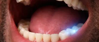
We only see 40% of the tooth (the crown part), and the remaining 60% is hidden under the gum. To “see” the hidden part, X-rays are used.
Today, many modern dental centers offer this service, and in most cases, the equipment available is enough to get your hands on a detailed clinical picture. However, before implantation or complex surgery, you may need more information – in these cases, it is better to contact a specialized diagnostic center that has modern high-precision equipment at its disposal.
The procedure is performed in a special room where the floors and walls are covered with lead to protect neighboring offices. Before beginning, the patient is asked to remove all metal jewelry, as their presence at the time of diagnosis can greatly distort the final results. Next, a protective lead apron must be worn to reduce radiation exposure to the body. If we are talking about targeted imaging, a special sensor connected to the x-ray unit is fixed on the causal tooth. The specialist presses a button and the machine starts working.
Computed tomography is performed somewhat differently. In this case, the patient’s head and chin are fixed using a special stand, and once the machine is started, the X-ray tube with a sensor begins to rotate around the head, making a whole series of pictures literally in half a minute – then a three-dimensional model of the patient’s jaw is formed on the computer.
They pass through tissues of different density, forming a corresponding imprint on the film:
- Fillings, artificial crowns and other dense substances block X-rays, so they appear as white objects;
- soft tissues, various fluids, as well as natural enamel and dentin are reflected in different shades of gray;
- inflamed areas and cavities will be shown as dark spots.
The procedure itself is performed in a separate office and lasts no more than a minute. The patient wears a protective lead apron and approaches the machine. Then you need to put a piece of film to the sick tooth and breathe quietly through your nose. The doctor will do the rest.
How the teeth and jaws look on the x-ray image
The result of the examination is an image of the tooth, a separate section, or the entire dentoalveolar system, which can be transferred to film or simply saved digitally. Trying to diagnose problems with such an image on your own is practically useless, because only an experienced specialist will be able to understand and correctly decipher the image.
The cost of X-rays in dentistry
It is quite difficult to give a precise answer about how much an x-ray of the teeth will cost – it all depends on the radiography method, the extent of the area under study, the type of equipment used, and the pricing policy of the medical institution. The table below shows approximate prices for different types of X-rays.
| INTRAORAL X-RAYS TYPE | AVERAGE COST | COST RANCE |
|---|---|---|
| Bitewing | $35 | $25-$50 |
| Periapical | $35 | $25-$50 |
| FMX | $150 | $100-$300 |
| Occlusal | $50 | $25-$100 |
| EXTRAORAL X-RAYS TYPE | AVERAGE COST | COST RANCE |
|---|---|---|
| Panoramic | $130 | $100-$250 |
| Ceph | $150 | $70-$300 |
| Cone-beam CT | $350 | $150-$750 |
Standard costs*:
- Although traditional x-rays record the image on film, lots of dental experts are changing to digital x-rays, which sends images to a computer. Whether x-rays use film or are digital does not appear to affect the price, which is typically based on the kind of x-ray taken– bitewing, periapical, scenic or occlusal. Rates differ by location and by private dental practitioner.
- Bitewing x-rays, which show the upper and lower back teeth, are often taken in sets of two (right and left) for children or four for adults and can cost $11-$28 for a single bitewing, or $18-$105 a set. They demonstrate how the teeth line up together and whether there’s any decay in between the teeth or bone loss due to severe gum disease. Bitewing x-rays are typically taken during a routine checkup or teeth cleansing, to try to find tooth decay.
- Anticipate to pay $14-$33 for a periapical x-ray, which is similar to a bitewing but shows more of an entire tooth from root to crown plus any supporting teeth, to determine dental issues below the gum line or in the jaw, like an affected tooth, an abscess, a cyst or a tumor. A full-mouth series of x-rays can cost $80-$260 and is normally done during a first check out to a dental expert. The series can consist of 14-21 separate images, often 4 bitewings and 10-17 periapical x-rays.
- Likewise called a breathtaking radiograph, a breathtaking x-ray can cost $55-$160. It offers a broad view of the jaws, mouth, teeth, sinuses and nasal areas, to highlight issues like affected teeth; bone irregularities; cysts, growths or other developments; infections; and fractures. A panoramic x-ray might be included in a total package price for braces.
- An occlusal x-ray (likewise called a palatal x-ray) reveals the roof or floor of the mouth, and generally costs $18-$42 each. Not as common as bitewing or breathtaking x-rays, occlusal x-rays are used to display extra teeth or teeth that have actually not grown through the gum; jaw fractures; a cleft palate; foreign things in the mouth or developments such as a cyst or abscess.
A separate fee may be charged for printing the image on film and having it transcribed by a specialist in writing. As for where to take x-rays, it should be noted that most modern dental centers are equipped with the necessary radiology equipment to obtain sight and panoramic images.
Another issue is if you have to undergo serious treatment, surgery and especially single-step implantation. Here it is very important to anticipate all the nuances and carefully examine the condition of the jawbone, to select the right places for implants. It will require a cone beam or multispiral tomography, and for this, experts recommend that you go to specialized institutions with high-quality and high-precision equipment.
*according to costhelper.com