What are abnormalities of shape and size of teeth? We will discuss the causes, diagnosis, and treatment methods in this article.
Definition of the disease
Anomalies of shape and size of teeth are variants of the development of teeth and the dental system, deviating from the norm.
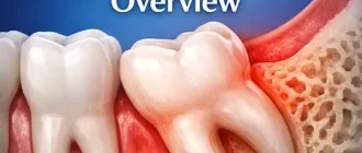
Malformations of the maxillary system are divided into large (congenital) and small. Congenital anomalies include facial clefts such as the cleft lip, cleft palate, etc., and minor anomalies of tooth eruption, number, shape, size and structure. Unlike congenital malformations, minor anomalies are not accompanied by significant violations and do not threaten the life of the patient, but they significantly affect the aesthetics of the tooth and tactics of treatment, and can lead to tooth decay, malocclusion and other complications.
Pro Tip: This Portable Water Flosser is up to 50% more effective than traditional floss and can save you hundreds on professional dental cleanings. Its 300ml tank and cordless design make it perfect for maintaining healthy gums. Check the current deal on Amazon.
This article describes frequently occurring small anomalies of tooth shape and size: macro- and microdentia, tooth fusion, etc. Absolute and relative indicators of their frequency and prevalence are shown in the table below.
| Types anomalies | Anomaly subtypes | Frequency of anomalies (%) | Prevalence anomalies (%) |
|---|---|---|---|
| Anomalies sizes | Macrodentia | 0,42 | 0,16 |
| Microdentistry | 7,87 | 3,08 | |
| Anomalies of shape | Tooth fusion | 0,21 | 0,08 |
| Taurodontism | 11,27 | 4,41 |
Subtypes of anomalies and causes of their development
All anomalies of tooth shape and development can be formed under the influence of negative factors that disturb the development of fetal tissues and organs during pregnancy. These include:
- medications;
- food additives;
- viruses;
- Industrial poisons;
- alcohol;
- Tobacco smoke, etc.
Macrodentia is characterized by an increase in the size of the teeth – a large crown and a tapering root. It can affect all or individual teeth. The prevalence of macrodentia of permanent teeth is 0.03-1.9%. More often this pathology occurs in men. The large crown size causes problems with tooth eruption and leads to dental deformities.
Microdentia is characterized by the reduction of one or all teeth. According to epidemiological studies, the prevalence of the anomaly ranges from 1.5% to 2% and is more common in women than in men. Teeth with this pathology usually have a cone shape.
Dental Care: This Rotating Electric Toothbrush removes up to 400% more plaque than a manual brush. With a built-in pressure sensor and 8 replacement heads included (a 2-year supply), it’s a cost-effective way to maintain a professional clean at home. Available on Amazon.
Macro- and microdentia can be hereditary in nature. Since the deposition of the follicles (rudiments) of permanent teeth occurs at different times, from the 5th month of intrauterine development to 3 years of age, the number of abnormal teeth will depend on the effects of negative factors during this period.
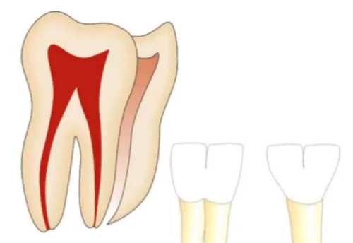
Taurodontism (bull’s tooth) refers to an abnormality of the tooth shape. It manifests as an enlarged pulp chamber, the “core” of the tooth, consisting of connective tissue, nerves, blood and lymph vessels. The base of the pulp is shifted closer to the apex of the tooth, there is no narrowing at the level of the cemento-enamel junction.
Taurodontism can occur at any age, but is more common in patients 13-19 years old. As a rule, the anomaly does not manifest itself before this age, since the roots of permanent masticatory teeth are formed before the age of 13.
Dental evagination (protrusion) affects the crown of the tooth. The abnormality occurs during the period when the rudiments of the teeth are being laid and formed. It appears as an additional cusp that protrudes from the crown of the tooth. Such a cusp consists of enamel and dentin. Its size, structure and location vary widely. It can be horny, conical or pyramidal in shape. Otherwise, this anomaly is called a cusp, claw cusp, Leong’s premolar, and occlusal enamel pearl.
The causes of the development of eugenichesia are not fully elucidated. The influence of X-linked and autosomal dominant inheritance has been suggested. In X-linked inheritance, the abnormality occurs only in men, and women are only carriers of the mutant gene. In autosomal dominant inheritance, boys and girls with dental intussusception are born equally often, and at least one parent of the child also has this anomaly.
Dental intussusception causes the enamel to fall inside the dental crown. Otherwise, this anomaly is called “tooth-in-tooth.” The invagination occurs before the calcification of the tooth tissue during intrauterine development. It can affect any tooth, but most commonly occurs on the upper lateral incisors (front teeth).
Symptoms of abnormal shape and size of teeth
In macrodentia, some teeth are enlarged, so that the remaining teeth do not have enough space in the dentition. Therefore, macrodentia is accompanied by deformation of dental arches and displacement of teeth.
In microdentia, one or more teeth are reduced, resulting in gaps between them.
Doubling and fusion of teeth usually cause complaints of aesthetic defects. These anomalies can only be distinguished from crowding of the teeth by X-rays.
In taurodontism, the tooth looks normal outwardly because its body and roots lie below the alveolar margin, i.e., they are hidden under the gingiva. Therefore, this anomaly is detected only by X-ray data.
Dental invaginations and evaginations are characterized by poor tooth aesthetics, contribute to plaque accumulation, food clogging and tooth decay. A dental intussusception can cause irritation and trauma to the tongue while speaking or chewing. It is sometimes accompanied by pain in the temporomandibular joint due to excessive pressure on the protruding tubercle.
Pathogenesis of tooth shape and size anomalies
The causes and mechanism of isolated macrodentia, in which only one or a few teeth are enlarged, are not known. Generalized macrodentia, affecting all teeth of the same jaw, is usually associated with systemic disorders or syndromes, such as insulin-resistant diabetes (type 2 diabetes), pituitary giantism, Costen syndrome, facial hemihyperplasia, KBG syndrome, Ekman-Westborg-Julin syndrome and chromosome (47, XYY) disomy syndrome. The pathogenesis of all these diseases is associated with abnormal development of organs and systems.
Generalized microdentia is rare and is part of syndromes such as pituitary nanism, Gorlin-Chaudhry-Moss syndrome and Williams syndrome (elf face syndrome). Their pathogenesis is also associated with abnormal development of organs and systems.
During tooth fusion, the periodontal ligament atrophy occurs, which causes the tooth to be positioned close to the jaw bone or another tooth. Due to the absence of interdental (interdental) bone and functional irritation of the periodontium, the layers of cementum between the roots of the teeth overlap and merge.
Some authors believe that the fusion of teeth is due to the tight fit of dental rudiments during the formation of dental tissues. Others attribute the appearance of this anomaly to the impact on the rudiments of a severe infection suffered in childhood.
Most experts believe that the fusion of teeth involves multiple dental rudiments or one but split, that is bifurcated. Splitted rudiments can either develop separately as normal and overcompleted, or fuse together at a certain stage of development.
If the fusion of tightly adjoining rudiments occurs during the formation of dental tissues, the fusion occurs after its completion. The cement of the teeth is involved in the fusion, and only the roots are fused. Due to the fusion of the dentin of both teeth, a single enamel and cementum are formed.
Taurodontism (bull tooth) develops when calcification of the pulp chamber is delayed during tooth formation. This anomaly can manifest itself in isolation. Sometimes it is associated with hypophosphatasia or abnormalities caused by the divergence of sex chromosomes: Klinefelter syndrome, Down syndrome, Martin-Bell syndrome, Mora syndrome, tricho-dento-cost syndrome and Maroteau-Lamy syndrome.
Dental evagination also occurs during the formation of the dentition. It is thought to be caused by the protrusion of the inner epithelium and papilla of the dental rudiment into the stellate reticulum, a group of cells located above the enamel nodule that disappear after the first layer of enamel has formed.
The pathogenesis of dental invagination is not fully known. Most authors believe that the cause of the anomaly lies in the curvature or cessation of enamel growth during its development. Moreover, the curvature of the enamel can be associated with the growth pressure of the dental arch, and the cessation of its growth is accompanied by the normal development of the surrounding tooth tissues.
- Classification and stages of development of anomalies of shape and size of teeth
- Macrodentia can be true or relative, localized (partial) or generalized (complete).
In true macrodentia, the abnormal teeth are significantly larger than the rest. In relative macrodentia, the size of the teeth is normal or slightly enlarged, and they are located on small, underdeveloped jaws.
In localized macrodentia, one or more teeth are slightly larger than the others, while the morphology of these teeth is unchanged. In generalized macrodentia, all teeth are larger than normal.
Macrodentia and microdentia of permanent teeth are divided into four groups:
- abnormality in the size of individual teeth;
- abnormality in the size of a group of teeth;
- abnormality in the size of teeth on one jaw;
- abnormality in the size of all teeth.
Dental intussusception is divided into three types:
- The first type – a small invagination covered by enamel is within the crown and does not extend beyond the boundaries of the enamel-cement junction;
- second type – the intussusception is covered by enamel, penetrates the root, but remains as a blind (separate) sac, sometimes associated with the pulp of the tooth;
- third type – the intussusception passes through the entire root, creating an additional hole in its apex or periodontal area (lateral wall of the root), with no connection to the pulp. It can be completely covered by enamel or cement.
Taurodontism is also subdivided into three types:
- Hypotaurodont – CB:R ratio ranges from 1.1 to 1.3;
- Mesotaurodontitis – CB:R ratio ranges from 1.3 to 2;
- hypertaurodont – CB:R ratio is greater than 2.
Complications of tooth shape and size anomalies
Because of the discrepancy between the size of the teeth and the shape of the jaw, microdentia and macrodentia lead to a variety of dental anomalies: the formation of gaps between the teeth (diastems and treble), crowding, misalignment, rotation of the teeth around their axis and bite problems.
Dental evagination is complicated by pathological erasure and fracture of the formed cusp. Later these damages can lead to necrosis (necrosis) of the pulp and the development of periapical infection, which subsequently becomes the cause of the destruction of the tooth.
The height of the evagination in the anterior group of teeth can reach the incisal edge of the crown. This anomaly affects not only the aesthetics, but also the occlusion of the teeth. All this can cause pain in the gingival and temporomandibular joint area.
Teeth with evagination usually have deep fissures – natural grooves in the enamel located on the chewing surface of the tooth. They contribute to plaque buildup, which makes teeth more susceptible to decay.
Invagination and taurodontism do not lead to complications, but because of their irregular structure create difficulties in treatment: canals of such teeth are quite difficult to be filled.
Diagnosis of anomalies of shape and size of teeth
To diagnose anomalies of the size of the teeth often used a technique developed by Doctor of Medicine Zubkova L.P. According to this method, the pathology is identified by measuring and summing the width of the four upper and lower incisors:
- with normal tooth size, the width of the upper incisors will be 28-32 mm, and the width of the lower incisors will be 22-24 mm;
- with relative macrodentia, the width of the upper incisors will be in the range of 33-34 mm, and the width of the lower incisors will be in the range of 25-27 mm;
- with absolute macrodentia, the width of the upper incisors will be more than 35 mm, and the width of the lower incisors will be more than 28 mm;
- in microdentia, the width of the upper incisors will be less than 28 mm, and the width of the lower incisors will be less than 22 mm.
Dental fusion and fusion almost always affect the front teeth, and fusion affects the second and third molars on the upper jaw. Examination and radiography are sufficient to make the diagnosis.
Taurodontism is detected only by X-ray. It is characterized by an enlarged pulp chamber in the shape of a rectangle. The body of the tooth is elongated, and the roots and root canals are shortened. At the same time, the size of the crown remains normal.
You need to distinguish from a hereditary tooth pathology in which the formation of dentin is disturbed. Such a tooth has a large pulp chamber and wide apical (root canal) openings on the X-ray. The roots themselves are not fully formed.
Dental evagination can be seen with the naked eye, but still requires radiographs. The examination allows to exclude concomitant dental anomalies: intussusception, overcompleted teeth, etc. In the picture, the eugenosis consists of normal enamel and dentin.
Treatment of anomalies of shape and size of teeth
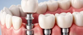
Treatment of macrodentistry consists of aesthetic rehabilitation with restorations or prosthetic manipulations: preparing the teeth, covering them with veneers or crowns.
With microdentia, orthodontic treatment and comprehensive rehabilitation are possible. Orthodontic treatment includes moving teeth and closing gaps, as well as creating the necessary space in the tooth row for subsequent dental rehabilitation with restorations or covering them with veneers or crowns.
To improve the aesthetics and function of a tooth with an anomaly, surgery, orthodontic treatment, pulp extraction, and dental restoration with veneers and crowns may be necessary. However, not all patients with tooth shape anomalies need treatment. It is not necessary if:
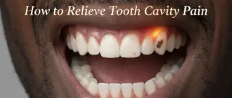
- There is no caries;
- The patient considers the aesthetic appearance of the teeth satisfactory;
- The occlusion of the teeth is not faulty, and this does not result in functional overloading of the individual teeth;
- The cusp in the euagination of the tooth is not sharp and does not erode.
To reduce the risk of unintentional fracture of the claw-like cusp located on the masticatory tooth, the evagination is bonded to the tooth surface using a composite (special cement). It is also possible to grind the tooth with a dental evagination, but after such a procedure, the tooth may become more sensitive to external stimuli.
Dental fusion can be suspected if the tooth does not move with the correct orthodontic load (e.g., when correcting a bite with braces) or if there is obvious resistance when the tooth is extracted. In these cases, treatment will consist of depulling the tooth (removal of the pulp) and surgical separation of the fused roots.
There is no specific treatment for taurodontism. However, in order to successfully treat the root canals of such a tooth, the peculiarities of its structure should be taken into account. During prosthetics of bovine tooth, it is recommended not to use pins, as such a tooth has no stability – it should not be used as a support.
Prognosis. Prevention
Prevention of dental anomalies is to lead a healthy lifestyle and eliminate the negative effects of external factors (drugs, food additives, viruses, industrial poisons, alcohol, tobacco smoke, etc.) during pregnancy.
Medical methods of prevention include genetic counseling and ultrasound diagnosis (USG). Genetic counseling is mandatory if relatives, parents or older children have birth defects. Ultrasound diagnosis should be conducted by appointment during the 10-13 weeks of pregnancy, as well as from 16 to 22 weeks and in the third trimester. With its help, up to 60-70% of abnormalities can be identified. Additionally, to diagnose malformations during pregnancy, amniotic fluid analysis, placenta biopsy, or biochemical analysis of the mother’s blood may be performed.
With abnormally shaped teeth it is important not to forget about the prevention of dental caries and its complications. For example, it is desirable to perform dental restorations, sealing or elimination of deep fissures when teeth are fused or fused.