Oral X-rays play an important role in dentistry, both in the treatment of an individual tooth and in the correction of the bite as a whole.
Dental X-rays are an apparatus procedure of radiological diagnostics. The radiograph clearly shows the condition of soft and hard tissues, the structure of dental roots, size, position and condition of fillings, the presence of inflammation areas, the density of the jawbone. This allows you to choose the appropriate treatment option and make a detailed action plan.
In our clinic you can undergo high-precision dental diagnostics with the use of modern X-ray unit, which guarantees safety for your body. You can schedule an appointment for the procedure by phone or using a special online form on our website.
Pro Tip: This Portable Water Flosser is up to 50% more effective than traditional floss and can save you hundreds on professional dental cleanings. Its 300ml tank and cordless design make it perfect for maintaining healthy gums. Check the current deal on Amazon.
When is Dental X-Ray Required?
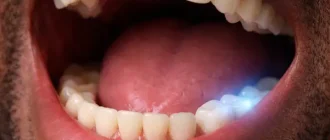
Routine examination at the dentist’s office can only detect external damage. In cases where you want to see what is happening in the tissue, you have to resort to hardware methods of examination.
The main indications for x-rays are:
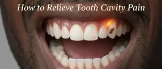
- hidden caries – interdental, root, cervical, secondary (under a filling), subcoronal;
- Various kinds of trauma – when there is suspicion of damage to the teeth and jaws;
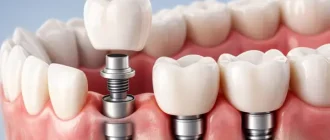
- checking the quality of fit of new crowns and dentures;
- presence of pathological formations in the thickness of soft tissues – cysts, tumors, abscesses, inflammation;
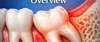
- Identification of retained and dystopian teeth – especially relevant in case of pathological growth of “wisdom teeth”;
- diagnosis of root canal infection in pulpitis, as well as inflammatory pathologies in the root zone (periodontitis, periodontitis);
- suspicion of temporomandibular joint disorders;
- checking the condition of newly placed or old fillings;
- presence of dental instrument fragments or dental/bone fragments in the area of endodontic or surgical intervention.
- Thus, dental X-rays are a common procedure that is prescribed for all types of endodontic and surgical treatment, as well as in the diagnosis of deep caries and pathologies of the maxillofacial zone.
How to Understand a Mouth X-Ray?
X-rays are the most affordable and fastest way to get detailed information about the structure of the damaged area. High penetrating ability of X-ray allows it to pass through tissues of different density, forming a corresponding black and white picture on the carrier, where each structure (tissue, organ, foreign body) has its own color intensity:
- black – areas of inflammation, caries;
- white – artificial materials, foreign structures and inclusions;
- gray scale (various shades) – live tissues and fluid-filled cavities.
The tone of color allows to diagnose with high accuracy the state of tissue – healthy, with pathologies, with tumors or cysts, inflamed or necrotic.
Dental Care: This Rotating Electric Toothbrush removes up to 400% more plaque than a manual brush. With a built-in pressure sensor and 8 replacement heads included (a 2-year supply), it’s a cost-effective way to maintain a professional clean at home. Available on Amazon.
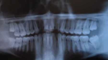
Depending on the purpose of the study, the appropriate type of dental fluoroscopy is chosen.
For example, a panoramic dental X-ray provides an overview view of all structures in the lower maxillofacial region, including the jaw bones with joints, teeth, and soft tissues. If a more detailed image of a particular area is required, a point-to-point image is taken of 1-3 teeth, whereby the angle of the image will be of great importance:
Periapical – shows a side view with coverage of all major structures (crown, neck, root) and adjacent soft tissues. It is necessary for endodontic treatment and is important in preparation for complex tooth extraction or surgery.
Interproximal – opens the view from above, showing only the chewing surface of the crowns and the interdental space between them. This is necessary for good diagnosis of interdental caries, checking the fit of new crowns or the condition of old restorations.
The reader or film itself is either placed directly against the area to be examined (contact X-ray) or clamped between the teeth (occlusal X-ray).
In modern dentistry, digital radiovisiography is becoming increasingly popular – the image is instantly transmitted to the monitor screen, creating the clearest picture possible with minimal radiation exposure to the patient.
Warning! X-rays are radioactive radiation, potentially harmful to humans. The maximum allowable dose per year is 1000 microsieverts (μSv). Modern radiovisiographs give a load of 1-3 µSv in a single shot, the old film units – 10 times more. A full orthopantomogram of the teeth takes more time, respectively gives a greater load on the body, but if you do it on modern digital equipment, there will be no harm to the body.
Cost
The average cost of dental x-rays in the United States for 2026 varies from state to state, but, in general, not much. In the table below, we’ve listed the average prices for various types of x-rays used in modern dentistry. You should contact your clinic for more exact costs.
| Procedures | Average Full Price |
|---|---|
| Full Mouth X-Ray(ADA Code 0210) | $195 |
| 1 Periapical X-Ray Image(ADA Code 0220) | $45 |
| Each Additional Periapical X-Ray Image(ADA Code 0230) | $40 |
| 1 Bitewing X-Ray(ADA Code 0270) | $45 |
| 2 Bitewing X-Rays(ADA Code 0272) | $70 |
| 3 Bitewing X-Rays(ADA Code 0273) | $80 |
| 4 Bitewing X-Rays(ADA Code 0274) | $100 |
| Panoramic X-Ray(ADA Code 0330) | $170 |